Peer Reviewed
ECG education
Left bundle branch block
Abstract
An ECG of a healthy young man shows a left bundle branch block. What are the potential causes and significance of this ECG pattern?
Key Points
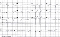
- Generally, in patients with LBBB, the QRS complex is more than 120 msec in duration with upright monomorphic R waves in V5, V6, aVL and lead I without Q waves. There is no secondary R wave inV1.
- Incomplete LBBB is diagnosed when a patient has the ECG criteria for LBBB but the QRS duration is less than 120 msec.
- It is difficult to be definite of an acute myocardial infarction in the presence of an LBBB without a rise in the serum troponin level and/or classic symptoms and signs.
- Patients with incomplete or complete LBBB require full investigation, including a cardiac echocardiography, assessment of cardiovascular risk factors, consideration of functional stress testing and a cardiology consultation.
- Patients with no impairment of exercise tolerance and no evidence of cardiac failure do not require any treatment other than reducing their cardiovascular risk factors as appropriate.
- Pacemaker cardiac resynchronisation therapy is required for patients with LBBB who have a reduced ejection fraction, those who are symptomatic with exertion and those who have cardiomyopathy.
Get full access
Buy this article
Single article purchases are temporarily unavailable due to site maintenance.
If you would like to purchase an article during this time, please email us at [email protected] with the article details and we'll assist you directly. We'll also let you know when online purchasing is available again.
Thank you for your patience and understanding.
Already a subscriber? Login here.