Peer Reviewed
ECG education
P wave abnormalities
Abstract
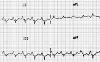
Yosef, aged 75 years, is a lifelong smoker. He has enjoyed good health for many years, but has experienced progressive shortness of breath, which has recently been more severe. He has never had pneumonia but has had intermittent bronchitis. He has been diagnosed in the past with emphysema, but has not been able to stop smoking. His only medications are combination fluticasone and salmeterol 250 microgram/25 microgram, one inhalation twice a day. Yosef has decided to have a transurethral prostate resection and the anaesthetist has requested (among other tests) a routine ECG. This has been performed and the result is shown in Figure 1.
Key Points
- P waves are most prominent, and therefore most easily seen, in leads II, III, aVF and V1.
- Each P wave should be less than 120 msec in duration (length; equivalent to three small squares) and under 2.5 mm in amplitude (height) in the limb leads and under 1.5 mm in amplitude in the precordial leads.
- P waves are normally upright in leads II, III and aVF, biphasic in lead V1 and inverted in lead aVR.
- P waves are absent when there is no conduction from the sinoatrial node to the atrium (sinoatrial block or arrest) and in some types of atrioventricular nodal rhythms.
Get full access
Buy this article
Single article purchases are temporarily unavailable due to site maintenance.
If you would like to purchase an article during this time, please email us at [email protected] with the article details and we'll assist you directly. We'll also let you know when online purchasing is available again.
Thank you for your patience and understanding.
Already a subscriber? Login here.